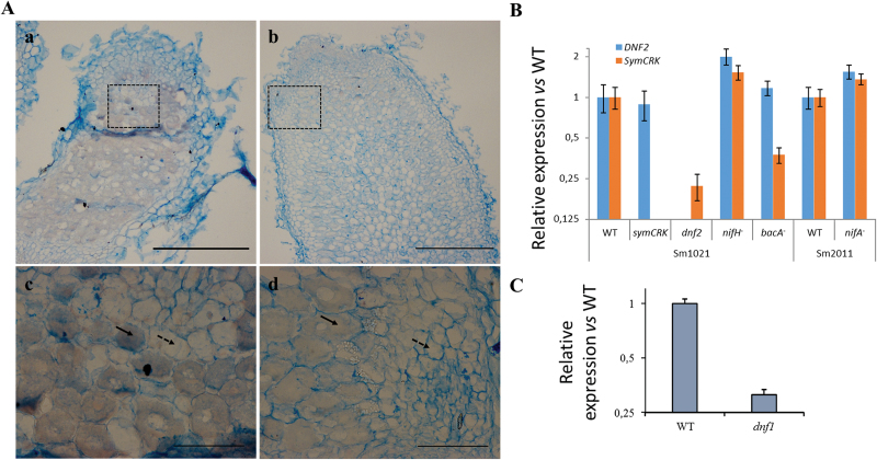
Fig. 3.
SymCRK is expressed in infected cells. (A) Expression of SymCRK was investigated using in situ hybridization and (B, C) RT-qPCR. Using antisense probe (a, c), signal was detected in infected cells (indicated with plain arrows) of the nitrogen fixation zone and not in uninfected cells (indicated with dashed arrows); sense probe was used as a control of specificity (b, d). c, d represent magnification of the zones delimited by a dashed rectangle in a and b. Scale bars represent 300 and 30 µm in whole nodule sections and in enlargement respectively. (B) RT-qPCR analyses revealed that SymCRK expression is strongly reduced in nodules of the dnf2 mutant. In contrast, the expression of DNF2 is not altered in the symCRK nodules. Also, expression of SymCRK was strongly reduced in the bacA induced nodules (B) as well as in the 14dpi nodules of the dnf1 mutant (C). B, C: error bars represent standard errors of three biological experiments with two technical replicates.

