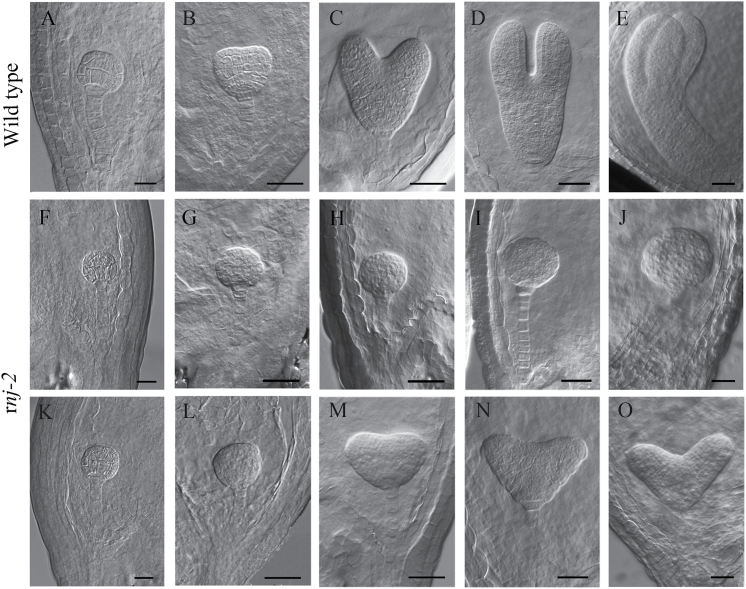
Fig. 2.
Embryogenesis in wild-type and rnj-2/+ plants examined by differential interference contrast microscopy. (A–E) Embryos from the globular stage to the bent cotyledon stage in wild-type ovules: (A) globular stage; (B) transition stage; (C) heart stage; (D) torpedo stage; (E) bent cotyledon stage. (F–J) The irregular globular rnj embryos from the same siliques at the different development stages as in (A–E). (K–O) The rnj embryos with abnormal cotyledons from the same siliques at the different development stages as in (A–E). Scale bars = 20 μm.

