
Fig. 3.

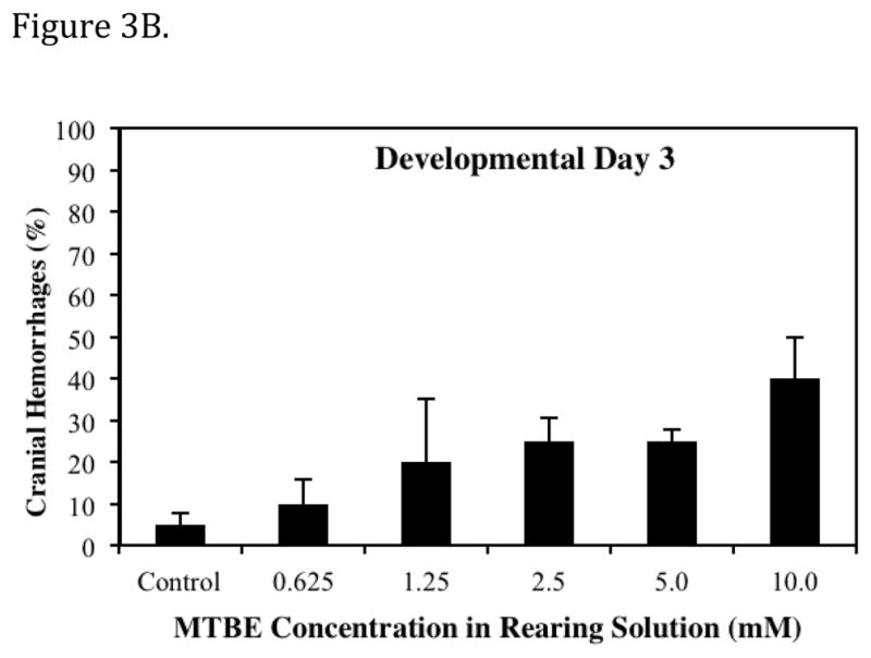

Dose dependent effects of MTBE on the percent of embryos exhibiting a specific vascular lesion. (A) Developmental Day 2 begins with the High Pec stage of embryo development. At this stage the CCV is a broad vein across the yolk. With MTBE treatment, blood cells pool in the lower portion of the CCV. The percent of embryos exhibiting this lesion is the higher on day 2 than on other days. (B) Developmental Day 3 begins with the Long Pec stage of embryo development. At this stage the microvasculature of the brain becomes patent. Embryos exposed to MTBE trend toward a dose dependent increase in cranial hemorrhages. (C) By Developmental Day 4, most of the vasculature in the embryo is formed and patent. MTBE treated embryos exhibit a dose dependent increase in abnormal development of ISVs, located between the dorsal somite muscles. *Significantly different from control (P ≤ 0.05). Data represents the average of three independent MTBE dose response vial studies.