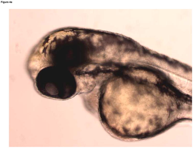
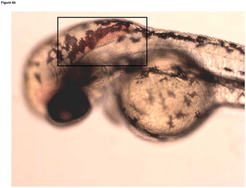
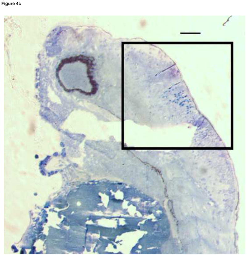
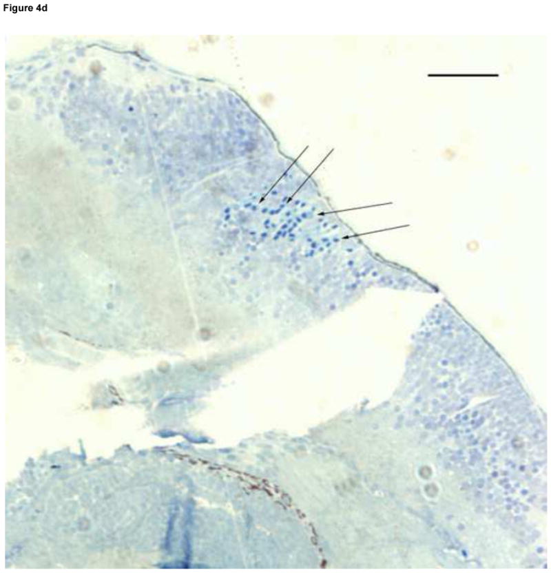
Fig. 4.


Developmental Day 3 (Long Pec) embryos with (A) normal cranial vascular flow or (B) a cranial hemorrhages in a zebrafish exposed to 10 mM MTBE. Cranial hemorrhages are predominantly present in the forebrain/midbrain region of the head, and the ventricles (10x); black box. (C) Toluiduine blue staining in a 72 hpf embryo revealed nucleated RBCs extravasated into the brain tissue (10x); black box. (D) Red blood cells did not appear to be undergoing hemolysis (20x); arrows. Scale bar = 100μm.
