Fig. 5.


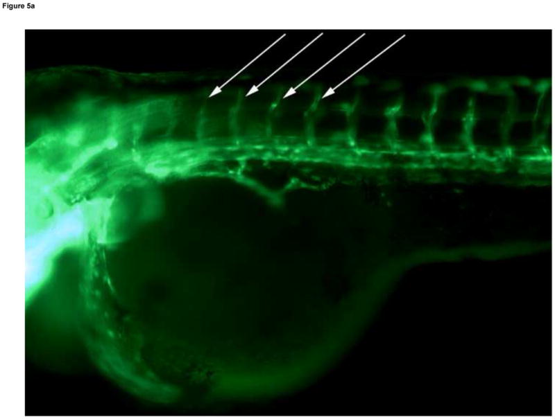
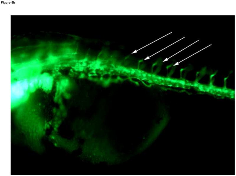
Developmental Day 4 (Protruding Mouth) embryos with (A) fully patent ISVs that appear as bright green edged tubes with a dim green lumenal area in controls (10x) - white arrows; or (B) abnormal ISV which lack proper circulation and appear as bright green vessels lacking a dim lumen in 10 mM MTBE treated embryos (10x) - white arrows. (C) Toluidine blue staining of day 4 control embryos reveals chevron shaped somite muscles; light blue stained tissue above dark blue yolk (20x). (D) Somites in MTBE treated embryos appear rounder and broader than in controls (20x) and (E) muscle fibers appear more disorganized (40x) – black arrows. Scale bar = 100μm.
