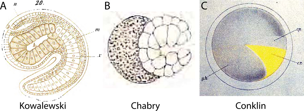
Figure 3. Historical approaches.
A) Hand-drawn image of a Ciona mid-tailbud embryo by Kowaleski (Kowalewski, 1866). The image shows 22 notochord cells instead of 40, and presents an overly regularized view of cell shape in the sensory vesicle and trunk endoderm, but the overall impression of the embryo’s chordate body plan is remarkably true to life. B) Hand-drawn image by Chabry of relatively normal gastrulation in an Ascidiella aspersa embryo where one of the two blastomeres had been killed with a fine needle at the two-cell stage (Chabry, 1887). C) Hand-drawn image by Conklin of the distribution of the yellow crescent material in the Styela egg that plays a key role in determining muscle cell fate (Conklin, 1905a).

