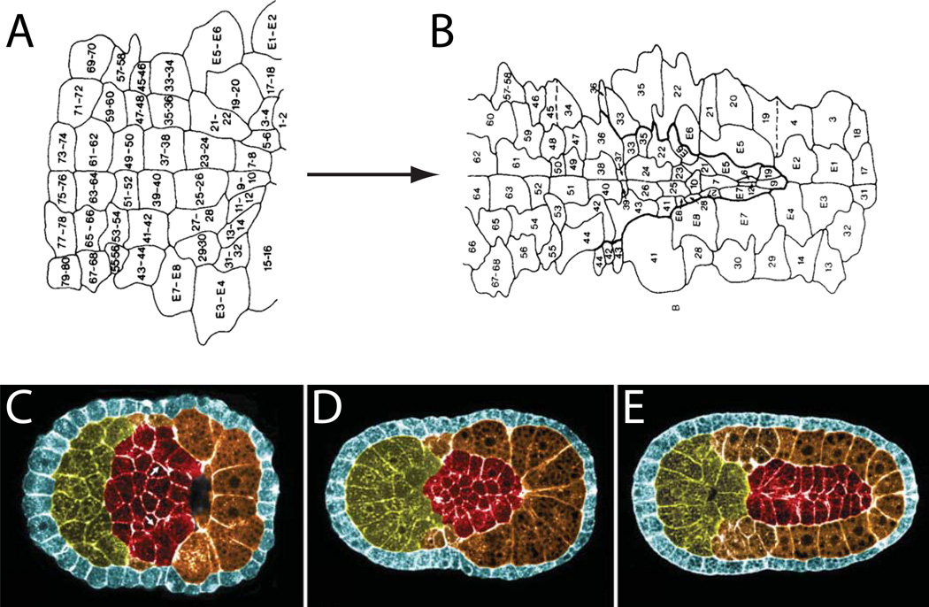
Figure 4. Early in toto approaches to neural plate and notochord development.
A,B) An early fate map of the Ciona neural plate through to early neural tube closure derived from histological sectioning and scanning EM of closely spaced timepoints of fixed embryos (Nicol and Meinertzhagen, 1988b). The blastomere numbering system used in these images is now obsolete, but the fundamental insight that the Ciona neural tube could be fate mapped with single cell resolution remains powerful. C,D,E) Pseudocolored confocal images of successive stages of notochord morphogenesis in Boltenia villosa (Munro and Odell, 2002b). These were among the first images to demonstrate how the entire ascidian embryo could be imaged by confocal microscopy to visualize fine details of cell and tissue morphology.

