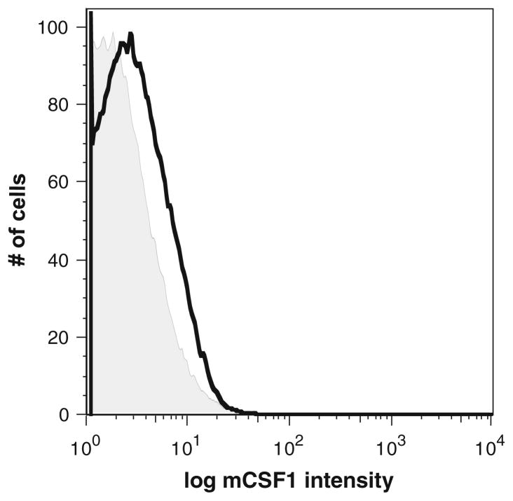
Fig. 4.
Flow cytometric analysis demonstrating the absence of mCSF1 protein expression in macrophages isolated from the k/o mice. Macrophages prepared from k/o mice (tinted) or from wt littermates (black) were analyzed by flow cytometry using a polyclonal antibody to CSF1. There is no staining of the k/o cells

