Figure 1.

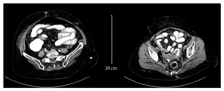
CT of the abdomen, showing extravasation of luminal contrast from the rectosigmoid region into a collection in the pelvis and air bubbles around the anastomosis. There is associated free air in the peritoneum.

CT of the abdomen, showing extravasation of luminal contrast from the rectosigmoid region into a collection in the pelvis and air bubbles around the anastomosis. There is associated free air in the peritoneum.