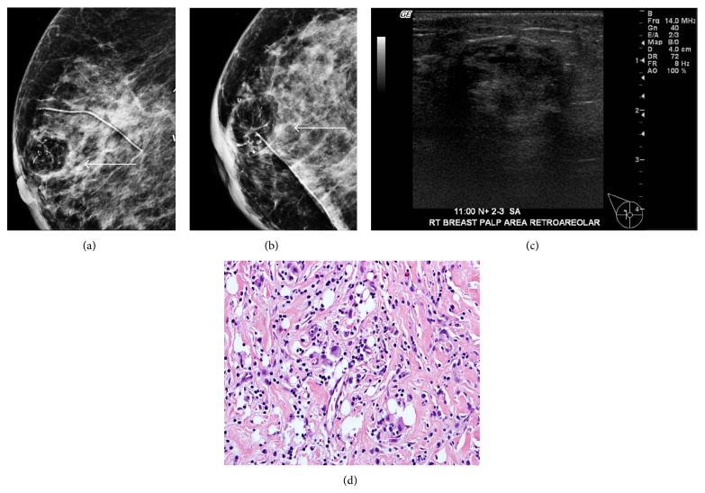
Figure 4.
85-year-old female with history of right breast mucinous carcinoma, lobular carcinoma in situ (LCIS), and ductal carcinoma in situ (DCIS) status after lumpectomy and radiation. Right breast mammogram ((a) and (b)) craniocaudal and mediolateral oblique views demonstrate a radiolucent round mass with dystrophic calcifications (arrow) (c). Targeted ultrasound demonstrates a heterogeneous hypoechoic mass with areas of posterior acoustic shadowing. The biopsy ((d); H&E, 400x) demonstrating dense fibrotic tissue with mixed chronic inflammatory infiltrate and scattered foamy histiocytes.

