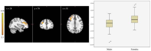
Figure 3.

Left image shows amygdala-frontal functional connectivity in which males show greater negative functional connectivity compared to females. The coordinates represent the position of the voxel with the highest intensity in MNI standard space (z-stat = 4.46); Right image compares the range of functional connectivity z-scores between the amygdala and these regions for the two groups.
