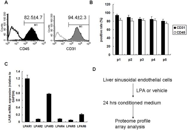
Fig 1. Liver sinusoidal endothelial cell isolation and LPA receptor expression patterns.
(A) Liver sinusoidal endothelial cells were isolated from mouse liver tissue and their purity was determined by CD45 (left panel) and CD31 (right panel) positives rate by flow cytometry analysis. (B) Liver sinusoidal endothelial cell purity during five serial passages. (C) LPA receptors’ mRNA expression determined by qRT-PCR. LPAR1 and LPAR6 mRNA expression was normalized to that of GAPDH mRNA expression (n = 3). (D) Strategy for investigating LPA effects on liver sinusoidal endothelial cells using proteome profile arrays.

