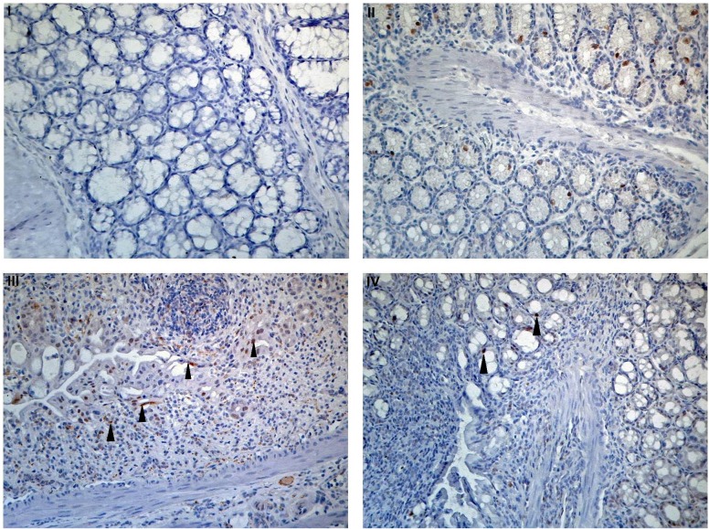
Fig 5. Apoptotic cell treatment inhibits NF-κB in DSS-induced colitis.
Mouse colon tissue sections were stained by immunohistochemistry assay using an antibody against mouse phospho-NF-κB (pNF-κB) p65. After immunostaining, slides were counterstained by hematoxylin. Images show pNF-κB p65 staining. All images are x200. (I) Untreated colon stained with HRP-anti rabbit secondary antibody only, without anti-NF-κB. (II) pNF-κB p65 staining in untreated colon. (III) Large pNF-κB p65 positive stain (black arrow) in 3% DSS-treated colon (3% DSS+PBS). (IV) Fewer pNF-κB p65 positive stain (black arrow) in 3% DSS treated colon with apoptotic cell infusion (3% DSS+ApoCell).

