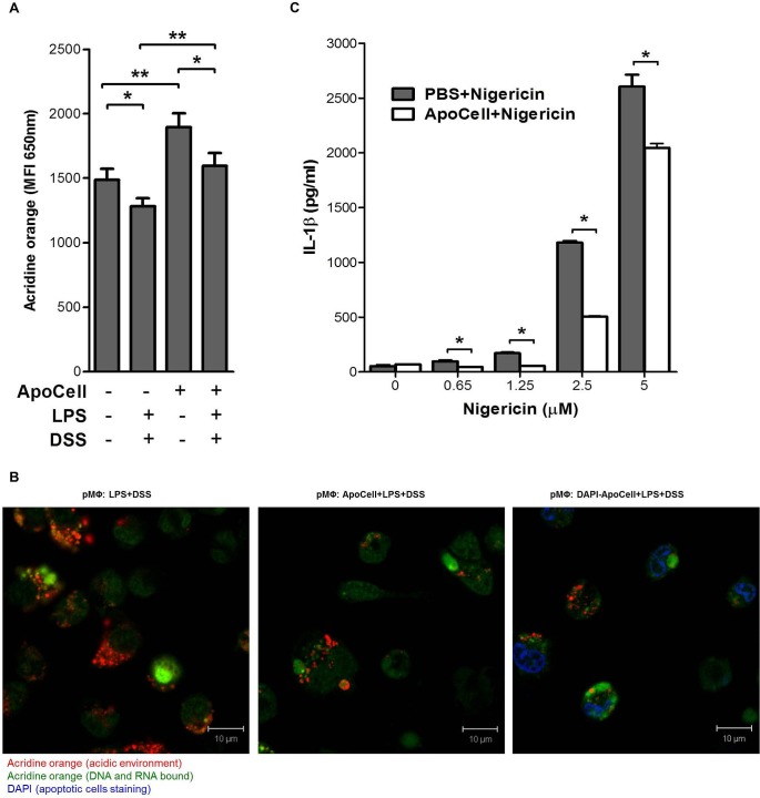
Fig 7. Lysosomal damage and K+ efflux in pMΦ.
(A) Flow-cytometer analysis of B6 pMΦ treated for 2h with apoptotic cells and/or 24h with DSS were stained with fluorochrome acridine orange (AO). Loss of fluorescence, which correlates with reduced numbers of lysosomes, was analyzed by flow-cytometer, excluding dead cells base on FSC/SSC parameters. Shown are means ± SEM of 4 independent experiments (*p<0.05, **p<0.03, one way ANOVA). (B) confocal microscopy of LPS primed pMΦ incubated (or not; left) for 2h with apoptotic cells (middle) or DAPI-stained apoptotic cells (right), stained with 1μg/mL acridine orange for 15 min and then incubated for 2.5 h with 3% DSS. Representative data from four experiments. (C) Apoptotic cell treatment inhibits nigericin-induced IL-1β secretion. B6 pMΦ cells were treated with nigericin at the indicated concentrations in the presence of LPS priming, with or without apoptotic cell treatment (*p<0.01, unpaired t-test).

