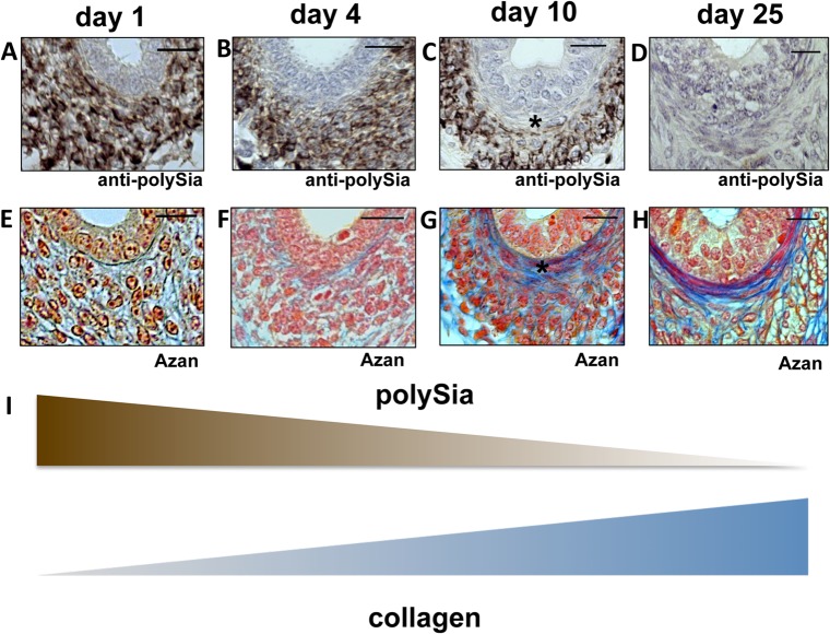
Fig 5. Immunohistological analysis of polySia+cells and collagen status of the inner longitudinal layer of smooth muscle cells in early and late postnatal mice epididymis.
Paraffin-embedded serial section of epididymis (cauda) at different time points (A-H). Sections of epididymis were stained with a mAb against polySia (A-D). For negative control, tissue sections were pretreated with endoN to degrade polySia (data not shown). An anti-SMA immunostaining was used to identify smooth muscle cells (data not shown). For collagen identification Azan staining was performed (E-H). * labels same positions of smooth muscle cells. Tissues were counterstained with Haemalaun (A-D). Scale bars representing 20 μm. (I) Illustration of the inverse correlation between polySia and collagen.

