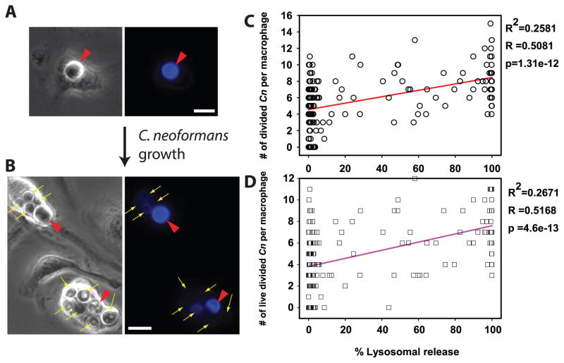
Figure 5. C. neoformans-mediated lysosome damage correlates with intracellular survival and replication of C. neoformans.
Fdx-loaded BMM were infected with Uvitex-2B stained Cn, and were assayed immediately or after 48h for lysosome damage and for the number, viability status and Uvitex-2B fluorescence signal of intracellular yeast. (A, B) Representative BMM, at (A) Baseline, brightly fluorescent undivided Cn (red arrow heads); (B) 48 h, when replication of the yeast diluted Uvitex-2B, permitting distinction between Cn that had divided (thin yellow arrows) and Cn that had not divided (red arrow heads). In A and B scale bars in lower right represent 10 μm. (C & D) Relationship between lysosome damage (x-axis) to the number per BMM of: (C) divided Cn, (circles) or (D) viable, divided Cn (squares). Correlation parameters for the illustrated data sets are listed to the right of each graph. Lysosome damage correlated with intracellular survival and proliferation of Cn. Data are combined from five coverslips across two independent experiments, totaling N > 170 BMM.

