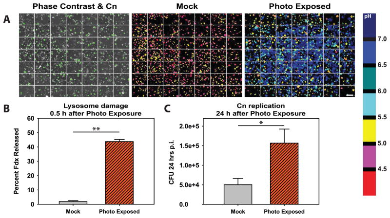
Figure 6. Photo exposure induced lysosome damage increases replication of C. neoformans.
BMM were loaded with Fdx and Texas Red dextran then infected with Uvitex-2B-stained Cn. One group of coverslips was photo exposed using bright orange/red light (“Photo Exposed”) while control coverslips were mock exposed (“Mock”). A grid array of images was then acquired of each coverslip using an automated acquisition sequence and the microscope motorized stage. (A) Representative images of coverslip imaging displaying 7 × 7 grids. Left panel displays phase contrast with Uvitex-2B staining overlayed in green to illustrate the presence of Cn. Middle and right panels display pH maps of a mock photo exposed coverslip (middle panel) and a photo exposed coverslip (right panel). The white scale bar in lower right of A represents 100 μm. (B) Average lysosome damage, quantified as the percent of Fdx released from the lysosome into the cytosol of the Cn-containing BMM. (C) Average CFU 24 post infection for coverslips photo exposed compared to mock exposed coverslips. In B & C data are combined from N > 5 coverslips combined from three independent experiments. * indicates p<0.05 and ** indicates p<0.005 student t test.

