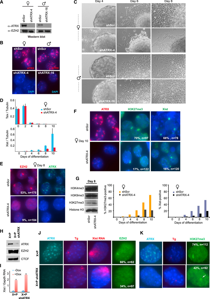
Figure 2. ATRX Is Required for Initiation of XCI during ES Cell Differentiation.
(A) Western blot of ATRX and EZH2 after stable ATRX KD.
(B) ATRX immunostaining after stable ATRX-KD.
(C) Representative phase contrast images of embryoid bodies during a differentiation time course.
(D) qRT-PCR analysis of Tsix and Xist RNA, normalized to tubulin RNA, during a differentiation time course. SE from three independent experiments shown.
(E) Immunostaining of EZH2 and ATRX at day 8 of differentiation, (n) and % with EZH2 Xi foci are indicated.
(F) Immunostaining of ATRX and H3K27me3 and Xist RNA FISH at day 10 of differentiation, (n) and % positive are shown.
(G) Left: western blot of indicated histone marks in day 8 ESCs. Graphs: Time course of Xist upregulation and acquisition of H3K27me3foci. n = 80–120 per time point.
(H) Western blot for ATRX, EZH2, and CTCF (control) in transgene (X+P) cells and in the same cell line expressing shATRX (X+P shATRX).
(I) qRT-PCR of Xist RNA before and after doxycycline induction in transgenic cells. SE from three independent experiments shown.
(J,K) Immunostaining of ATRX, EZH2 (J), and H3K27me3 (K), and Xist RNA/DNA FISH in indicated cells lines after 24 hr dox induction. % with shown pattern and sample size (n) are indicated.

