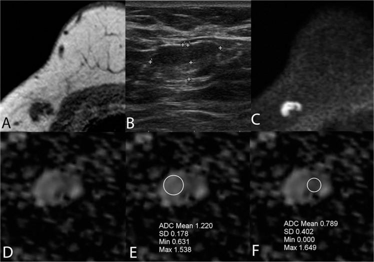
Fig 1. A false positive lymph node due to thickened cortex was true negative on DWI (A-F).
Imaging of a 41-year-old female with a BI-RADS 5 lesion in the right breast. An axillary LN with a cortical thickness of 3.9 mm was core biopsied. Final histology revealed a 30 mm, grade 3, T2 ductal carcinoma; the SLNB was benign. No recurrence occurred over a two-year follow-up. The LN was false positive on T1-weighted MRI (A) and US (B) due to thickened cortex, while DWI b = 800 (C,D) was true negative with ADC = 1.24 x 10⁻³ mm2/s (cut-off 0.812 x 10⁻³ mm2/s). The importance of correct positioning of the ROI and ROI’s information (including minimum value) is illustrated at the area of fatty hilar lobulation (E). False-positive ADCs can be obtained from the cortical area medially if the morphological images are not evaluated or the minimum value of 0 is accepted or is not available at the time of evaluation (F).

