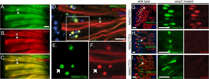
Fig 2. The proximal tail region of NINAC mediates RTP colocalization.
(A-C) Images obtained from fluorescent microscopy show that GFP-NINAC and RTP-RFP both localize to the photoreceptors’ rhabdomeric membranes (labeled R, arrow). (D-F) Confocal retina images of RTP-RFP and GFP-NINACTAIL localization with native NINAC expression. The boxed region in (D) is magnified for GFP-NINAC labeled in (E) and for RTP-RFP labeled in (F). One rhabdomere, labeled by RTP-RFP, is identified as “R” in D. The arrows in (D-F) identify cytoplasmic structures containing both RTP and NINACTAIL. (G-I) The GFP tagged tail constructs TD1, TD2 and TD3 expressed in cells with native NINAC, are found in proximity to the photoreceptor R1-6 nuclei (left column, DAPI staining labeled blue). RTP is found in the rhabdomere, and colocalizes only with the NINACTD1 construct (yellow). In middle and right columns, the GFP-tagged tail constructs TD1, TD2 and TD3 are coexpressed with RTP-RFP in a ninaC P235 null mutant background. RTP is found associated with the cytoplasmic structures containing NINACTD1, but is absent from cytoplasmic structures containing NINACTD2 or NINACTD3. Scale bars, 10 μm.

