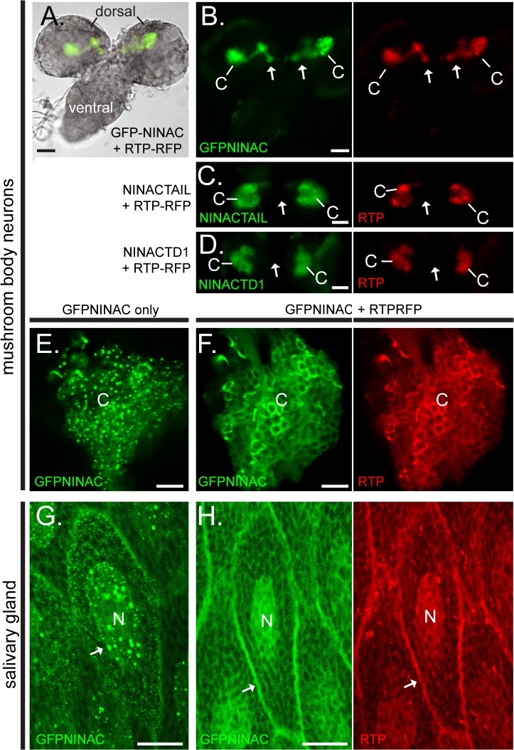
Fig 3. RTP and NINAC interact in multiple Drosophila cell types.
(A-F) The GFP-NINAC tail domain constructs were expressed with and without RTP-RFP in larval mushroom body neurons. (A) A bright field image of larval brain, overlaid with fluorescence images specifying GFP-NINAC and RTP-RFP location is shown. The dorsal and ventral brain regions are labeled. (B) Left and right panels are the fluorescent images from the larval brain shown in (A) detecting the location of GFP-NINAC and RTP-RFP respectively when both are expressed simultaneously. GFP-NINAC and RTP-RFP are colocalized within the Kenyon cell bodies (labeled C) as well as projections through the peduncle and into the neuronal lobes (arrows). (C-D) When RTP-RFP is expressed with NINACTAIL and—NINACTD1, both GFP-NINAC and RTP-RFP are localized only within the Kenyon cell bodies and not within the peduncle structures (arrow). (E,F) 1.5 μm confocal optical sections of the Kenyon cells (C) of the mushroom bodies expressing GFP-NINAC only (E) or GFP-NINAC and RTP-RFP (F). In the absence of RTP, the GFP-NINAC protein is found within punctate structures, while the presence of RTP-RFP prevents formation of GFP-NINAC puncta. (G,H) 1.5 μm confocal optical section showing GFP-NINAC only (G) or GFP-NINAC and RTP-RFP (H) localization in larval salivary gland cells. GFP-NINAC is found within punctate structures within the nucleus (labeled N), while expression of RTP-RFP prevents formation of these GFP-NINAC punctate structures and the two proteins are colocalized within the cytoplasm, likely associated with internal membranes (arrow). Scale bars, A-D: 50 μm, E,F: 10μm, G,H: 20 μm.

