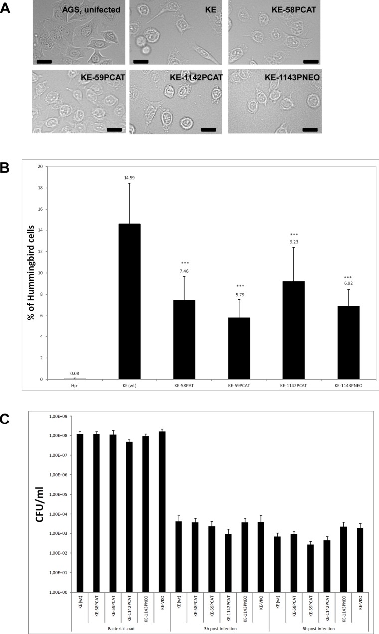
Fig 3. A) AGS cells co-incubated either with wild-type H. pylori cells (KE), ccrp deletion mutants as indicated or uninfected (AGS).
Co-incubation was performed at MOI of 100 for 4 h. Cells were visualized by phase-contrast microscopy (BZ-9000E (KEYENCE) microscope) to assess AGS cell morphology. Scale bar, 20 μm. B) Quantification of the percentage of elongated cells from (A). All samples were examined in triplicate in at least three independent experiments. Data are presented as mean value of three independent experiments. For each strain between 1830 and 2800 cells were counted and evaluated. Exact percentage values are indicated above the bars. Asterisks indicate a significant difference between the ccrp mutants and wild-type H. pylori (the P value was <0.001, as determined by Student's t test). C) Bacterial adherence analysis in AGS cells infected with KE88-3887 or ccrp deletion mutants as indicated. AGS cells were infected with H. pylori for 3 h and 6 h, respectively. The number of cfu per cell was determined as described in experimental procedures and normalized to ml.

