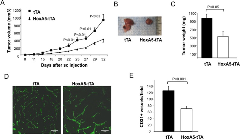
Fig 4. HoxA5 expression in EC inhibits angiogenesis and growth of allograph mammary tumors.
(A) Tumor volume in tTA (square) and HoxA5-tTA (triangle) 8 week old mice (3 weeks + Dox, 5 weeks—Dox), 32 days following subcutaneous injection of MMTV-PyMT tumor cells into syngeneic female FVB/n mice. The analysis revealed a significant reduction (p<0.01) in volume of tumors grown in HoxA5-tTA mice on days 25 through 32 (n = 5). (B) Micrograph showing representative tumors obtained 32 days after implantation of tumor cells into tTA (left) and HoxA5-tTA (right) mice. (C) Quantitative analysis of tumor weight 32 days after implantation into tTA or HoxA5-tTA mice. HoxA5-tTA mice showed a significant reduction (p<0.05) in tumor weight as compared to tTA mice (n = 5). (D) Immunofluoresence analysis of vascular density in tumors in tTA (left panel) or HoxA5-tTA (right panel). Vascular density was assessed by CD31+ staining of frozen OCT-embedded tissue sections of peri-tumor tissue from each animal. (E) Quantitative analysis of CD31+ vessels in tumors isolated at day 32 from HoxA5-tTA mice compared to those from tTA mice. HoxA5-tTA mice showed a significant reduction (P<0.01) in the number of vessels (n = 5).

