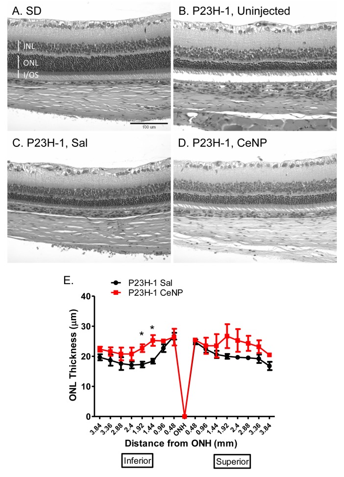
Fig 3. Morphometric analysis of ONL thickness in CeNPs- and saline- treated P23H-1 rats.
One μl of 1 mM CeNPs (172 ng) or saline was delivered to each eye of the animals at P23 and eyes were harvested at 28 dpi; three eyes from 3 individuals were examined per treatment group. (A-D) Representative photomicrographs of H&E stained retinal sections from wildtype, Sprague Dawley (SD) or P23H-1 animals uninjected or treated with either saline or CeNPs. Similar regions were shown: 1 mm from the ONH in the inferior region. (E) shows quantification of the ONL thickness measurements. The overall ONL thickness was higher in CeNPs-treated than in saline-treated animals although the increases were not statistically significant across many of the regions. INL = inner nuclear layer, ONL = outer nuclear layer, I/OS = rod inner/outer segment, ONH = optic nerve head. *P<0.05.

