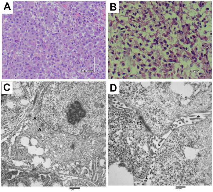
Figure 1. Images of malignant mesothelioma from patient tissue samples.
(A) Light microscopic image of H&E-stained right chest wall biopsy material showing large cells with clear to eosinophilic cytoplasm and enlarged nuclei with prominent nucleoli, typical of an epithelial mesothelioma. (B) Light microscopic image of H&E-stained right kidney tumor tissue from the autopsy. Cells are large with clear cytoplasm, suggestive of a renal cell carcinoma or a malignant mesothelioma. (C and D) Transmission electron photomicrographs of the right chest wall biopsy showing ultrastructural features of mesothelioma including perinuclear tonofilaments (^), fragmented, elongated microvilli (*), and giant desmosomes (⇧).

