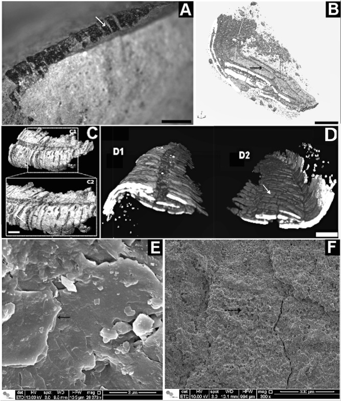
Fig 6. Corumbella werneri: ultrastructure of theca.
(A) GP1E-574a: three-dimensional specimen with theca. (B) 3D-rendered microCT of Corumbella theca,flipped by 180° compared to (A): interior view and detail for lamellae microfabric and plates (black arrow). (C) 3D-rendered microCT of Corumbella theca (A) without flipping in C1 and C2, with details of rings (white arrow). (D) compression and fragmentation along theca. D1 shows a transversal section in the fossil structure. Flipped by 180° of it produces D2, with details of small breakages (white arrow). (E) Details of lamellar plates by SEM (black arrow) and (F) pores in plates (black dashed arrow). Scale: 1mm.

