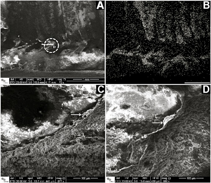
Fig 7. Corumbella werneri: ultrastructure of theca.
(A) SEM of a longitudinal section of a Corumbella theca. Dashed circle marks a detail of a sectioned ring. (B) EDS mapping of (A). Here it is possible to observe higher concentration o calcium in fragments of theca (represented by white dots) in comparison to the rock matrix and molds of fossil without fragments. (C) and (D) are details of the white dashed circle in (A), showing micro layers in Corumbella theca (white arrow). Scale for B: 1mm.

