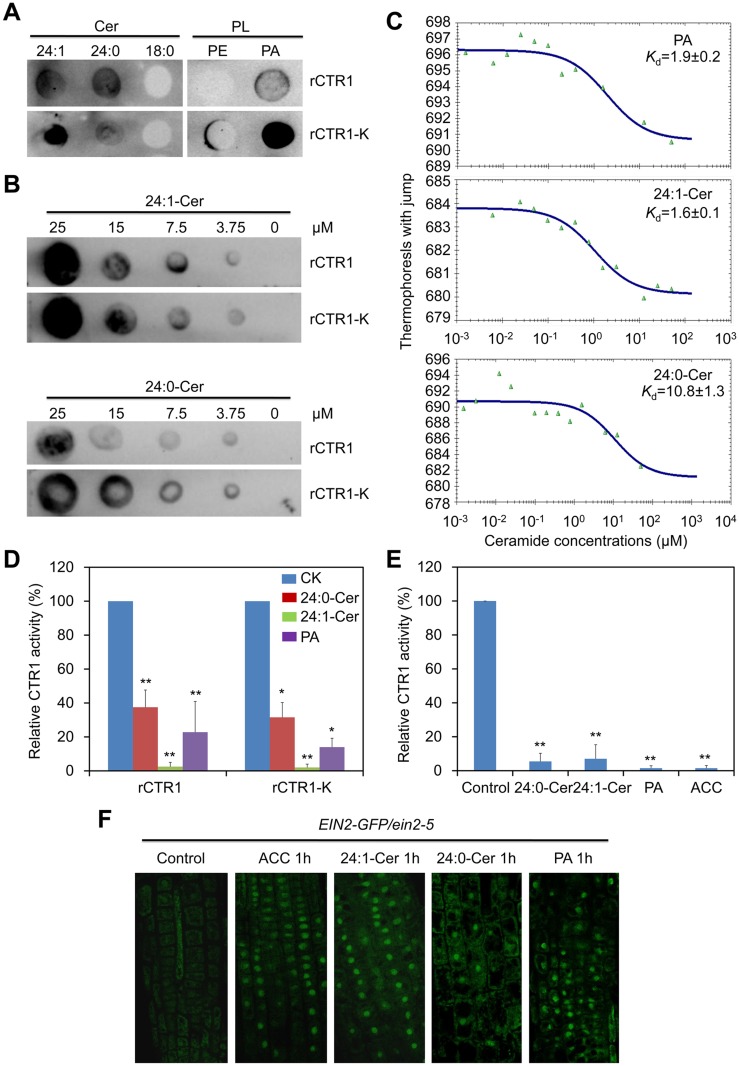
Fig 6. Ceramides interact with the kinase domain of CTR1 protein in vitro.
(A) Recombinant rCTR1 and CTR1 deletion mutant with kinase alone (rCTR1-K) binds ceramides/phospholipids on filters. Fifty micromole concentrations of various ceramides (24:1-Cer, 24:0-Cer and 18:0-Cer) and phospholipids (PE and PA) were spotted onto nitrocellulose and incubated with 1 μg/mL of either purified rCTR1/rCTR1-K protein. The binding was detected by immunoblotting using anti-GST antibodies and anti-His antibodies, respectively. (B) Binding of the rCTR1 and rCTR1-K to 24:1- and 24:0-Cer on filters. Various concentrations (0, 3.75, 7.5, 15 and 25 μM) of 24:1- and 24:0-Cer spotted onto nitrocellulose. (C) MST analysis of the interaction between rCTR1 and liposomes of PA, 24:1-Cer as well as 24:0-Cer. (D) Measurement of CTR1 kinase activity in vitro. The purified proteins (rCTR1 and rCTR1-K) were pre-incubated without or with 1 nM 24:0-ceramide, 24:1-ceramide and PA liposomes and subsequently used for kinase activity assay. The relative TR-FRET signals were calculated based on the fluorescence emission ratio at 665/620 nm and presented by subtracting the signals of negative controls purely containing the substrate without ligands. The signals of rCTR1 or rCTR1-K pre-incubated without liposomes were set as 100% and the relative CTR1 activities in the proteins pre-incubated with 24:0-ceramide, 24:1-ceramide and PA liposomes were calculated accordingly. The experiment has been repeated and data are means ±SD (n = 10). *P<0.05, **P<0.01 by Student’s t-test. (E) Measurement of CTR1 kinase activity in vivo. The 10-d-old Arabidopsis seedlings (WT and ctr1-1) were non-treated or treated with 10 μM ACC, 50 μM 24:0-ceramide, 24:1-ceramide and PA liposomes for 1 h and their total proteins were extracted for kinase assay. The relative TR-FRET signal was calculated based on the fluorescence emission ratio at 665/620 nm and presented by subtracting the background signal in the ctr1-1 mutant from WT. The signals of non-treated proteins were set as 100% and the relative CTR1 activities in the proteins treated with 24:0-ceramide, 24:1-ceramide and PA liposomes as well as ACC were calculated accordingly. The experiment has been repeated and data are means ±SD (n = 10). **P<0.01 by Student’s t-test. (F) Activation of EIN2-GFP translocation from the ER to the nucleus by ceramides. Roots of 7-d-old EIN2-GFP/ein2-5 seedlings were used for detection of GFP fluorescence at 1 h after treatment with either 10 μM ACC, 50 μM 24:0-Cer, 24:1-Cer and PA liposomes.

