Fig. 2.

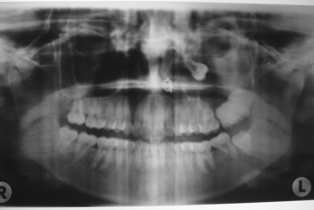
Preoperative panaromic radiograph showing a radiopaque lesion encircled by radiolucent band in the left maxillary sinus, 3rd molar near orbital floor, thickened sinus lining and complete bone loss distal to 2nd molar

Preoperative panaromic radiograph showing a radiopaque lesion encircled by radiolucent band in the left maxillary sinus, 3rd molar near orbital floor, thickened sinus lining and complete bone loss distal to 2nd molar