
Fig. 5.
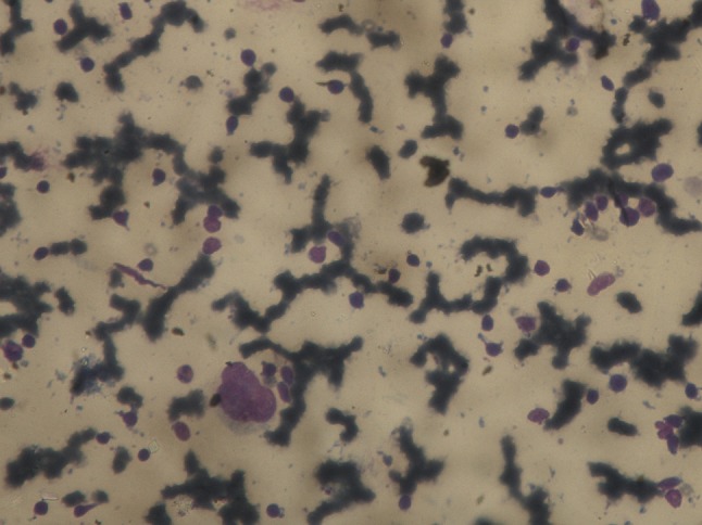
FNAC of the lesion showed numerous atypical cells scattered singly. Cells showed marked nuclear pleomorphism, binucleated and multinucleated forms (Reed Stenberg cells) along with multiple mitotic figures
Official websites use .gov
A
.gov website belongs to an official
government organization in the United States.
Secure .gov websites use HTTPS
A lock (
) or https:// means you've safely
connected to the .gov website. Share sensitive
information only on official, secure websites.
FNAC of the lesion showed numerous atypical cells scattered singly. Cells showed marked nuclear pleomorphism, binucleated and multinucleated forms (Reed Stenberg cells) along with multiple mitotic figures