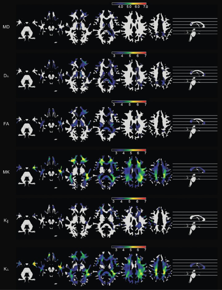
Figure 1.
Voxelwise maps of white matter abnormalities in patients with temporal lobe epilepsy compared with controls. The first and second rows demonstrate areas with an increase in MD and radial diffusion, respectively, in patients. The third row demonstrates areas with reduced FA in patients, while the fourth, fifth, and sixth rows demonstrate areas of reduced mean kurtosis, axial kurtosis, and radial kurtosis in patients, respectively. Reproduced with permission from (13). MD, mean diffusivity.

