
Figure 3.
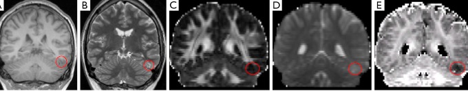
Focal cortical dysplasia in the left inferior temporal gyrus that is poorly defined on structural images including volumetric T1-weighted (A) and T2-weighted PROPELLER (B) and standard DTI images including FA (C) and MD (D) but easily visible as reduced ICVF (E). Reproduced with permission from (27). DTI, diffusion tensor imaging; FA, fractional anisotropy; MD, mean diffusivity; ICVF, intracellular volume fraction.