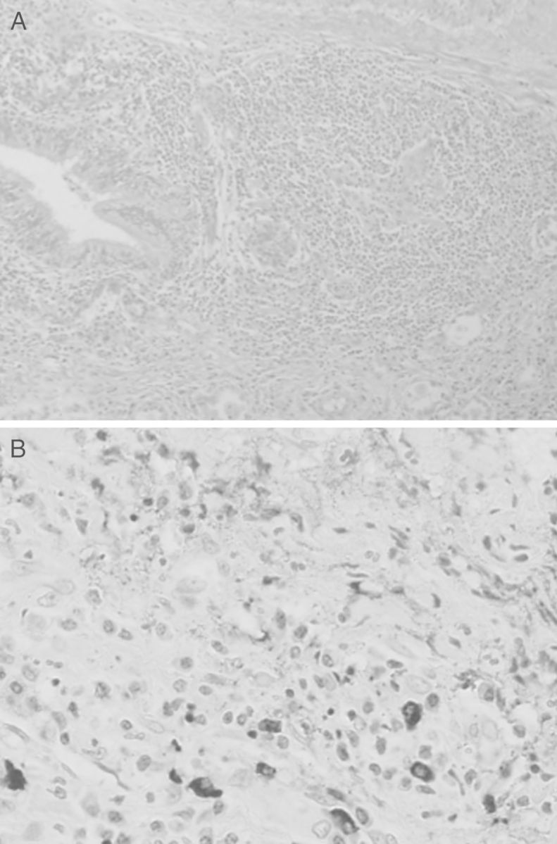
Fig. 5.

Light microscopy of the resected lung tumor shows heavy plasma cells and lymphocyte proliferation, and fibrotic changes of the interstitium. No malignant change is found (A). Immunohistochemistry microscopy shows IgG4-positive plasma cells in the specimen (B).
