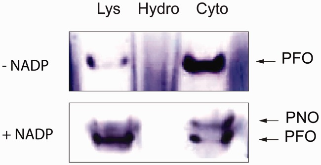
Fig. 2.

Histochemical detection of PFO and PNO in Mastigamoeba balamuthi cell fractions. Cell lysate (Lys) was separated into cytosolic (Cyto) and hydrogenosome-enriched (Hydro) fractions using differential centrifugation. Samples were separated under native conditions in a 6% polyacrylamide gel and stained for the oxidative decarboxylation of pyruvate in the presence and absence of NADP+ as an electron acceptor to distinguish PFO and PNO activity, respectively.
