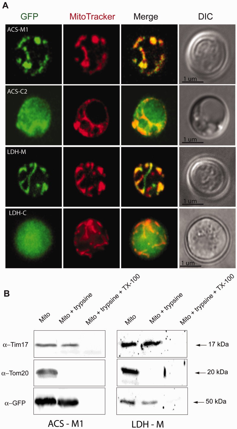
Fig. 3.
Expression and cell localization of Mastigamoeba balamuthi ACS and LDH in Saccharomyces cerevisiae. (A) Immunofluorescence microscopy. Mitochondrial (-M) and cytosolic (-C) versions of ACS and LDH were fused at the C-terminus with GFP and expressed in yeast. Mitochondria were visualized using MitoTracker. DIC, differential interference contrast. (B) Protease protection assay. Mitochondria were isolated from yeast expressing M. balamuthi ACS-M and LDH-M and treated with trypsin to remove proteins that are associated with the outer membrane; the mitochondria were treated with a combination of trypsin and the detergent Triton-X100 to disintegrate both membranes. Transblotted samples were probed using antibodies against Tom20 (an outer membrane marker), Tim17 (an inner membrane marker), and GFP, which is present at the C-terminus of ACS and LDH.

