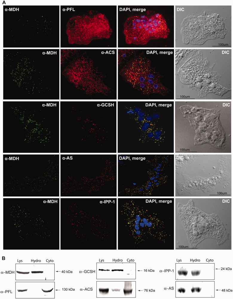
Fig. 4.
Cellular localization of PFL, ACS, AS, and IPP in Mastigamoeba balamuthi. (A) Immunofluorescence microscopy. Signals for GCSH, AS, and IPP clearly colocalized with hydrogenosomal MDH (PCC = 0.907 ± 0.023, n = 42; 0.902 ± 0.025, n = 41; and 0.876 ± 0.023, n = 38, respectively). ACS colocalized with MDH. However, some signal was observed in the cytosol (PCC = 0.659 ± 0.047, n = 43). Signal for PFL was consistent with its cytosolic localization (PCC = 0.411 ± 0.063, n = 39). Proteins were visualized using antibodies against human GCSH and M. balamuthi AS, ACS, IPP, PFL, and MDH. Alexa Fluor 488 donkey α-rabbit and 594 donkey α-mouse and α-rat were used as secondary antibodies. (B) Immunoblot analysis of cellular fractions. Cell lysate (Lys) was separated into cytosolic (Cyto) and hydrogenosome-enriched (Hydro) fractions using differential centrifugation and probed with the appropriate antibodies.

