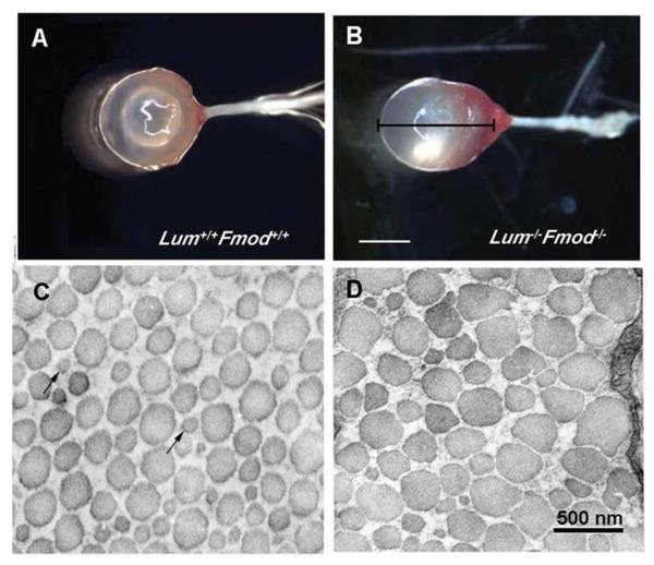
Figure 2.
The sclera of lumican/fibromodulin deficient mice. Photographs of whole eyes of wildtype mice (Lum+/+ Fmod +/+) (A) and lumican/fibromodulin deficient mice (Lum−/− Fmod−/−) (B), demonstrating significantly higher axial length of deficient mice as compared with wildytpe mice. Collagen fibril morphology in the posterior sclera of wildtype mice (C) and Lum−/− Fmod −/− deficient mice (D). Collagen fibrils from deficient mice displayed abnormal small-to very large-diameter fibrils with irregular contours. Bar in B = 2 mm; Bar in D = 500 nm. From: Chakravarti S., et al. Ocular and scleral alterations in gene-targeted lumican-fibromodulin double-null mice. Invest Ophthalmol Vis Sci 44: 2422–2432. 2003 Reproduced with permission © Association for Research in Vision and Ophthalmology.

