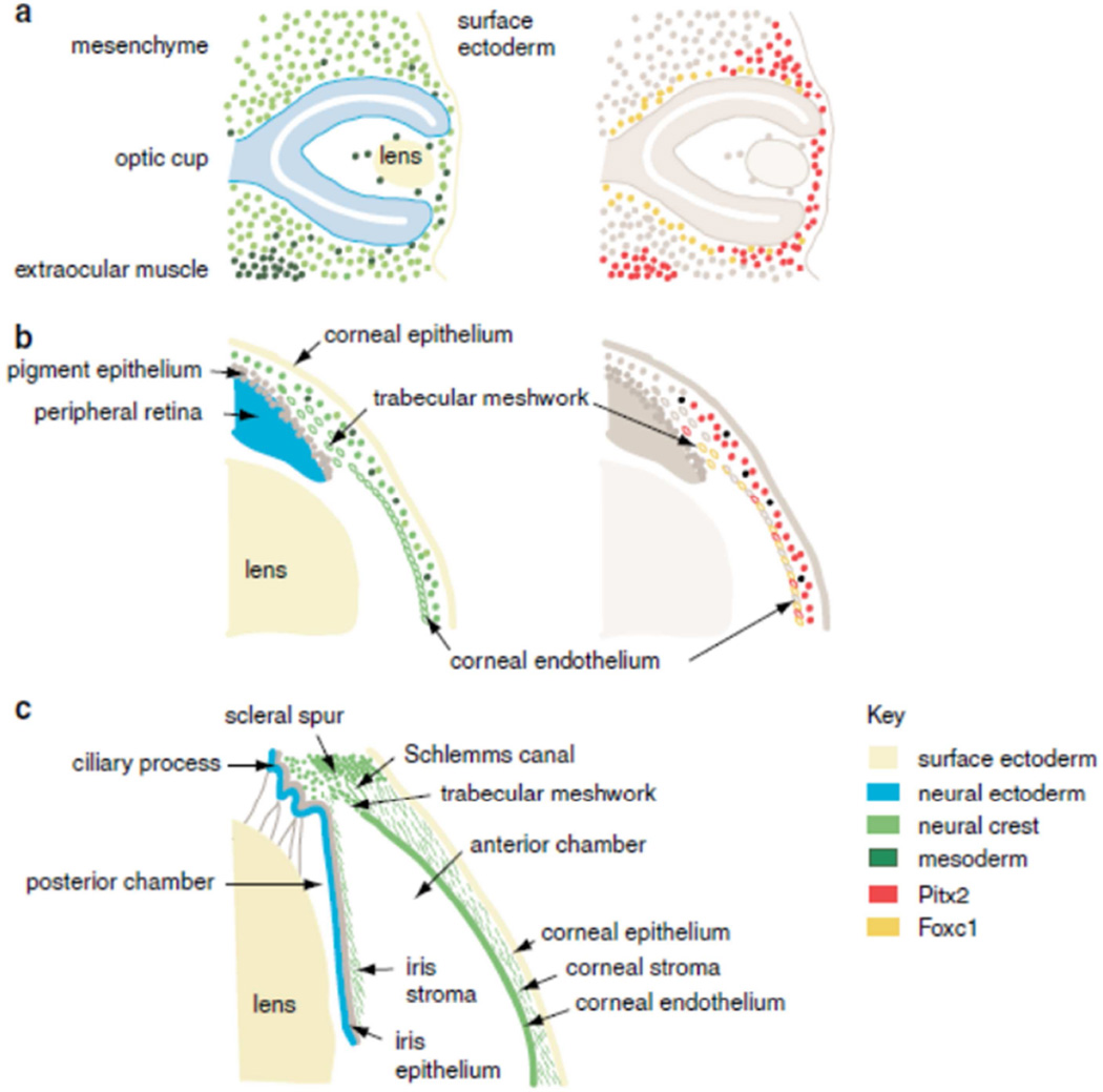
Figure 1. Development of the anterior segment of the eye.
(a) Optic cup stage, embryonic day 10.5 in the mouse equivalent to week 5 in human development. (b) Formation of anterior chamber, embryonic day 15.5 in the mouse equivalent to the 5th month of human gestation. (c) Mature anterior segment depicting the lens, iris, iridocorneal angle, the TM and the cornea. Key shows the color coding used to represent the embryonic origin of the anterior segment tissues in the right-hand plates, and the pattern of expression of the FOXC1 and PITX2 genes in the left-hand plates, based on published expression data. Reprinted with permission from (Sowden, 2007).

