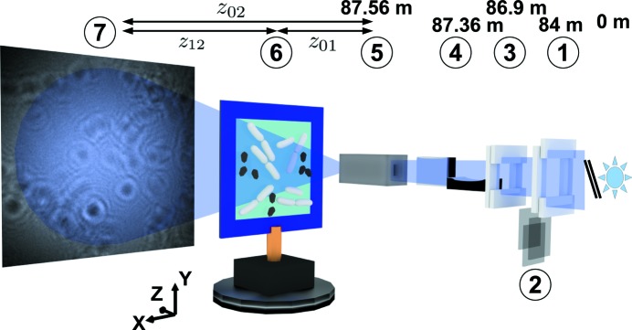
Figure 1.

Schematic of the GINIX waveguide setup: downstream of the undulator source and the monochromator (not shown): (1) slits S1, (2) attenuation foils made of Al, (3) slits S2, (4) KB mirror system, (5) X-ray waveguide (entrance in the focal plane of the KB mirrors), (6) bacterial sample at a distance  to the waveguide and (7) detection device at a distance
  to the waveguide. The flight tube between sample and detector is not shown in the image.
