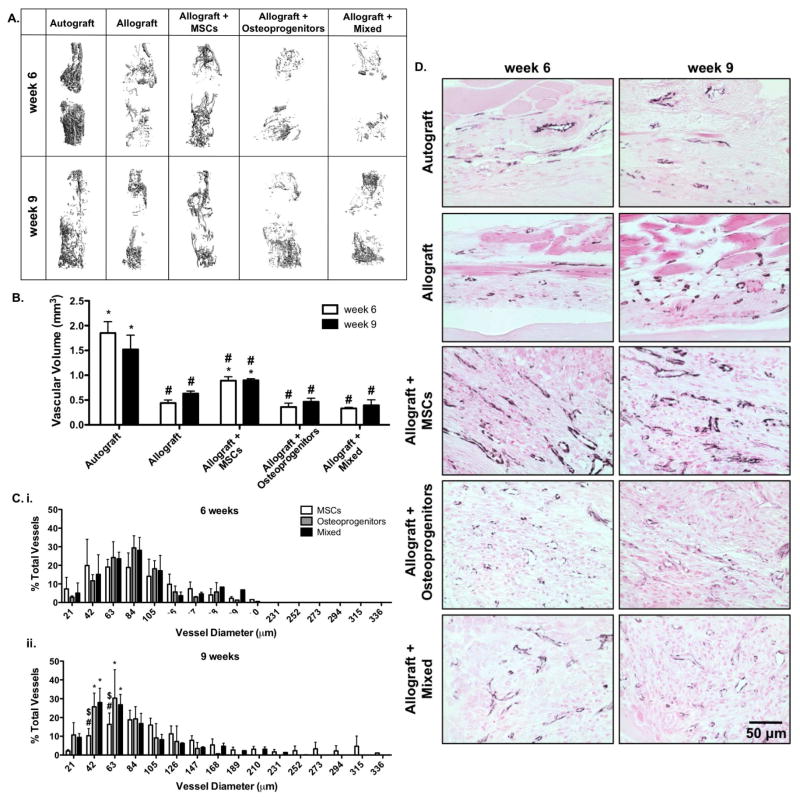
Figure 3.
Micro-computed tomography (μCT) scans taken 6 and 9 weeks post-implantation were reconstructed to assess in vivo host-mediated graft vascular infiltration (A). Subsequent quantification revealed that tissue engineered periosteum modified allografts transplanting a combination of MSCs and osteoprogenitors, or osteoprogenitors alone exhibited reduced vascular volume as compared to transplantation of MSCs alone (B) (n=6; error bars represent standard error of the mean; p-value of <0.05 indicates significance compared to allograft (*) or autograft (#)). This reduction in total vascular volume was shown to be due to decreased arteriogenesis and increased small vessel formation; as verified by micro-computed tomography thresholding (C) (n=6; error bars represent standard deviation; p-value of <0.05 indicates significance compared to MSCs (*), mixed (#), or osteoprogenitors ($)), and immunohistological staining for CD31 (D) (black; CD31, and pink; nuclear fast red counter stain and muscle).

