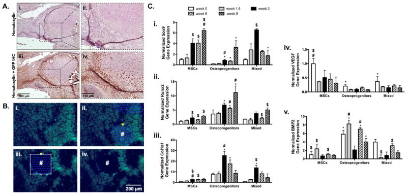
Figure 9.
Representative immunohistological staining for green fluorescent protein (GFP) showed limited incorporation of mixed (GFP+ mMSCs and osteoprogenitors) into the newly formed bone callus 9 weeks post-implantation (A). Despite this finding, fluorescence microscopy was used to locate transplanted mixed GFP+ mMSCs and osteoprogenitors within prepared histological sections (B) and laser capture microdissection (LCM) was performed to remove selected cells (#; Ci–Civ). Subsequent RNA isolation and gene expression analysis revealed that cells transplanted in a 50:50 ratio of MSCs to osteoprogenitors exhibited trends towards expedited endochondral ossification as determined by Sox9 (Ci.), Runx2 (Cii.), Col1a1 (Ciii.), VEGF (Civ), and BMP2 (Cv) gene expression (n=5–6; error bars represent standard error of the mean; p-value of <0.05 indicates significance compared to MSCs (*), 50:50 MSCs (#), or osteogenic MSCs ($)).

