Figure 2.
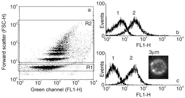
a) and b): Immunofluorescence against 1-μm lipid-coated colloids fused with wild-type baculovirions (wt-AcNPV). a) Dot plot, where each dot provides a measured event, for which the forward scattering (FSC-H) and the fluorescence in the FITC channel (FL1-H) were recorded and used for subsequent analysis. Region R1 is assigned to single colloids, and R2 to particle aggregates. b) Single colloids have been gated and their fluorescence versus particle counts are shown. Curve 1: Lipid-coated lbl colloids, curve 2: lipid-coated lbl colloids fused with wt-AcNPV. c) Inset: CLSM scan of a 3-μm colloid coated with AcNPV displaying an antibody-binding fragment of HIV-1 gp120 (curve 2). Curve 1: colloids with wt-AcNPV; curve 2: colloids displaying the HIV-1 gp120 epitope on their surface.

