
Figure 1.
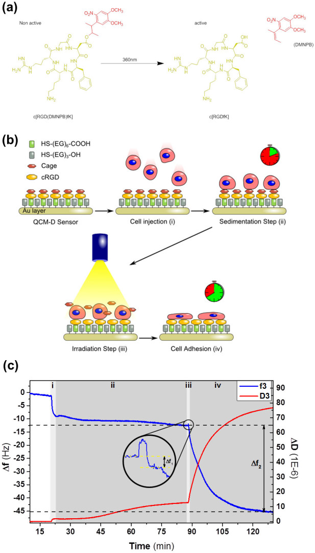
(a) Chemical structure of the photo-activatable adhesive peptide ligand c[RGD(DMNPB)fK] and the products generated upon photolysis. (b) Schematic of the steps to trigger synchronized cell adhesion and spreading. (c) QCM-D measurement of a integrin/RGD-mediated cell binding process on the c[RGD(DMNPB)fK] crystal. Changes in frequency f (blue line) and dissipation D (red line) through the experiment described in Figure 1b. i- cell seeding; ii- cell sedimentation; iii- UV irradiation through the window of the QCM-D chamber for RGD peptide activation (magnified inset); iv- integrin binding and cell spreading.