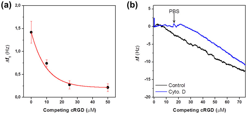
Figure 3. Integrin binding and not spreading is responsible for Δf1.
(a) Binding experiments were performed in the presence of increasing concentrations (0 to 50 μM) of competing soluble RGDfK peptide ligands resulting in monotonic decrease of Δf1. (b) Cells treated with Cyt. D were no longer able to spread after irradiation at minute 1 (blue line), as observed from the unchanged frequency, but were capable of accomplishing firm initial attachment since they remained immobilized on the substrate upon rinsing the chamber with PBS (minute 15). When adding Cyt. D-free PBS again, the cells regained their spreading properties again comparable to control cells (black line). Microscopy images confirmed these results by showing cells in a rounded shape in the presence of Cyt. D, and the usual flattening after a system rinsing with PBS (pictures not shown).

