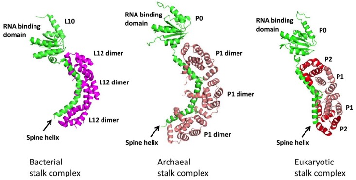
Figure 1.
Structural organization of bacterial, archaeal and eukaryotic ribosomes. Structures of bacterial, archaeal stalk complex were determined by X-ray crystallography [11,17], while the structural model of eukaryotic stalk complex was predicted as described [27]. Bacterial stalk complex is consisted of L10 (green) and 2 to 3 copies of L12 dimers (magenta), while archaeal stalk complex is consisted of P0 (green) and 3 copies of P1 homodimers (salmon). On the other hand, eukaryotic stalk is consisted of P0 (green), P1 (salmon) and P2 (red) in 1:2:2 stoichiometry. Two copies of P1/P2 heterodimers bind to the spine-helices of P0, presumably adopting a P2/P1:P1/P2 topology [19,24].

