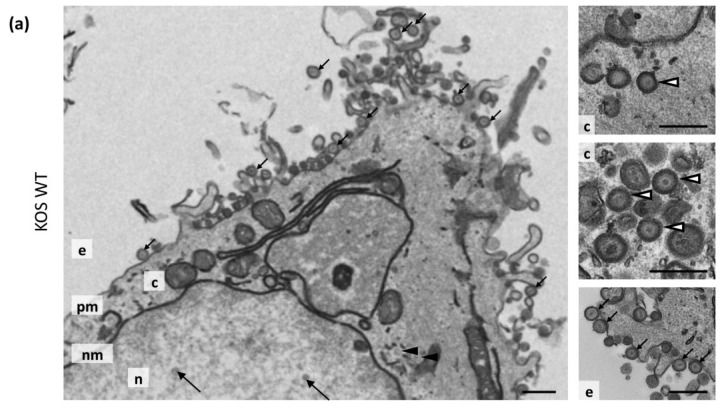
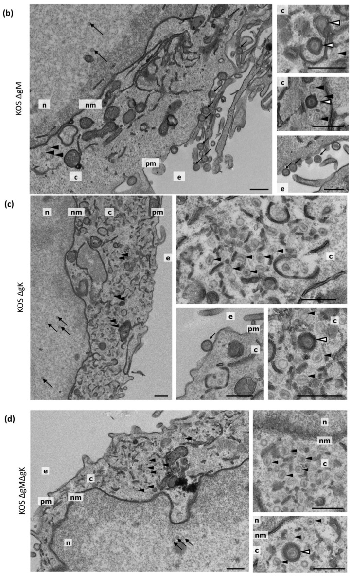
Figure 5.
Ultrastructural morphology of cells infected with KOS WT, ∆gM, ∆gK and ∆gM∆gK viruses. HaCaT cells were infected with (a) KOS WT; (b) ∆gM; (c) ∆gK; and (d) ∆gM∆gK virus at an MOI of 5 PFU/cell and processed for electron microscopy at 20 hpi. Representative nuclear capsids are marked with large black arrows, extracellular virions with small black arrows, cytoplasmic enveloped virions with white arrowheads and cytoplasmic naked capsids with black arrowheads. The nucleus (n), cytoplasm (c), nuclear membrane (nm) plasma membrane (pm) and extracellular space (e) are marked. Scale bars: 500 nm.


