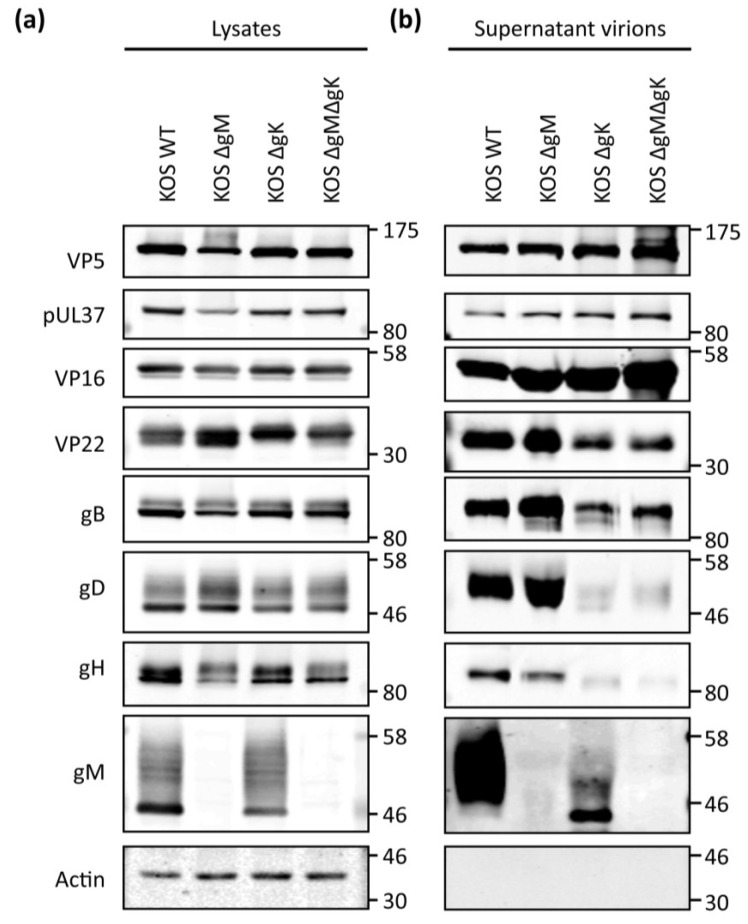
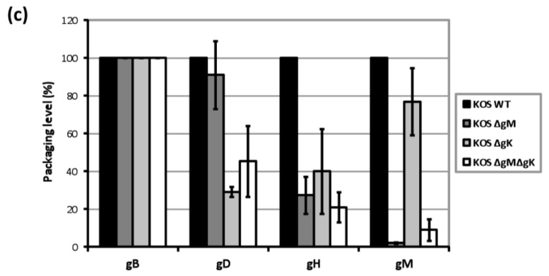
Figure 6.
Viral protein incorporation in WT, ΔgM, ΔgK and ΔgMΔgK virions. HaCaT cells were infected at 5 PFU/cell and after 24 hpi, (a) cell lysates were prepared and (b) progeny viral particles were isolated from clarified cell culture media by centrifugation through a 33% sucrose cushion. Cell lysates and supernatant-derived virions were separated by SDS-PAGE and analysed using the indicated antibodies. Molecular mass markers (in kDa) are indicated on the right; (c) Using densitometry, packaging levels were calculated by dividing the amount of protein detected in supernatant-derived virions (normalized for gB) by the amount of protein in supernatant-derived virions of WT virus (normalized for gB). Error bars represent standard error of three independent experiments.


