Figure 5.

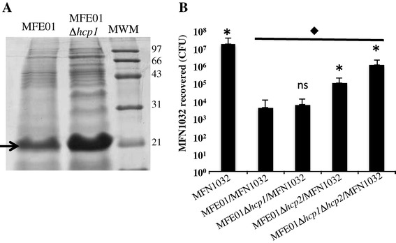
Hcp secretion and killing activity of P. fluorescens MFE01 and derivatives strains. A: Concentrated supernatants of MFE01 and MFE01Δhcp1 cultures were analysed by SDS-PAGE and Coomassie staining. Bands with a molecular mass similar to that of an Hcp protein (≈20 kDa), indicated by the arrow, were observed at a growth temperature of 28°C. MWM: molecular weight markers are indicated. B: Quantitative co-culture assays were performed. Prey cells (MFN1032 carrying pSMC21-gfp) were or were not mixed at ratio of 1:5 with P. fluorescens MFE01, MFE01Δhcp1, MFE01Δhcp 1Δhcp2 and MFE01Δhcp2; after 4 h at 28°C, MFN1032 cfu were counted (n = 4, the error bars represent standard error of the mean). * Indicates a significant difference in MFN1032 cfu (p-value <0.05) relative to the MFN1032/MFE01 assay; ns means no significant difference. ♦ indicates a significant difference in MFN1032 cfu (p-value <0.05) relative to the MFN1032 alone control assay.
