
Figure 7.
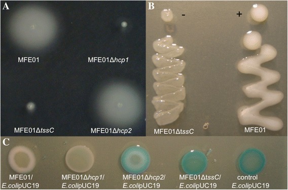
Phenotypes of the MFE01∆tssC mutant. A: Swimming assay on LB in 0.3% agar at 28°C during 24 h. MFE01 and MFE01Δhcp2 were motile contrary to MFE01Δhcp1 and MFE01ΔtssC strains (n = 3). B: Representative images of mucoidy of MFE01 and MFE01ΔtssC on LB agar at 28°C, 24 h (n = 3). +: mucoid, −: non mucoid. C: Effects of P. fluorescens MFE01 and its mutants on competitive E.coli on solid medium. Co-cultures were performed on LB medium supplemented with X-Gal (40 μg/ml) at 28°C during 24 h. Prey cells (E.coli DH5αmcr containing pUC19) were or were not mixed with P. fluorescens MFE01 or its derivatives at ratio 1:1. Blue is due to X-Gal degradation by E.colipUC19. The images shown are representative of three assays.