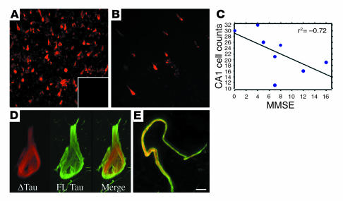
Figure 3.
ΔTau is found predominantly within AD brain and is inversely correlated with cognitive function. Large numbers of ΔTau-immunoreactive neurons (red) were observed in the hippocampus and entorhinal cortex of AD brains (A). In contrast, only occasional ΔTau-immunoreactive cells were observed in age-matched control cases (B). Preadsorption of the α-ΔTau antibody with immunizing peptide resulted in no immunofluorescence (A, inset). Regression analysis of the number of hippocampal CA1 cells labeled with α-ΔTau versus AD Mini-Mental State Examination (MMSE) scores revealed a significant inverse correlation between ΔTau and cognitive function (P = 0.05, r2 = –0.72; (C). High-power confocal microscopy demonstrated distinct subcellular localization of ΔTau (red) and the C-terminal–specific antibody T46 (green) within cell bodies (D) and dystrophic neurites (E). Scale bar: 65 μm (A and B), 8 μm (D), 5 μm (E).

